The Psychopath Whisperer: The science of those without conscience is printed in black and white. Here we provide color copies of the important figures in the book.
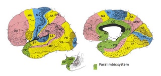
Figure 6. page 167. Summary of the brain regions believed to be implicated in psychopaths by the year 2000. Regions include the amygdala, hippocampal complex, anterior and posterior cingulate, anterior temporal pole, and the orbital frontal cortex. The numbers represent different areas of the brain as defined by the work of Dr. Brodmann. See prior figures for complete details on illustrations.
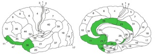
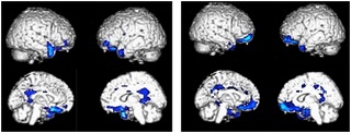
Figure 8. page 213. Results from the first structural MRI analyses of criminal psychopaths collected on the mobile MRI system. The colored regions depict areas of the brain that are atrophied in adult male criminal psychopaths. The areas represent the majority of the paralimbic system of the brain.
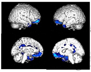
Figure 9. page 215. Results from the first structural MRI analyses of incarcerated youth collected on the mobile MRI system (left image). The colored regions depict areas of the brain that are atrophied in youth with elevated callous/unemotional traits. The results from adult males (right image) are shown for comparison. The results from the two groups are strikingly similar. In both groups the majority of the paralimbic system is showing reduced gray matter density or atrophy.